
Moltkiopsis ciliata / الحماط
Lithospermum callosum Vahl.
Salih, Halem, Halamah, Hamat, Ghabsha, Ealan
Callous-leaved gromwell
Halam, Hamat
Boraginaceae
Aerial parts
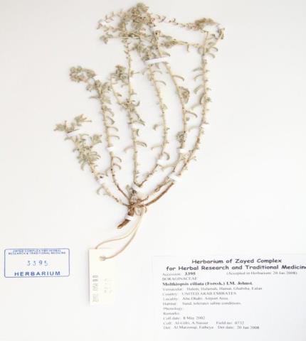
Herbarium specimen
Ethnobotanical Characteristics
Description
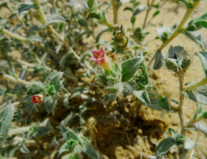
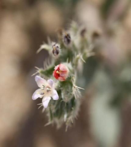
Small perennial herb up to 25cm with white younger branches, very smooth, and dark brown bark on older plants. Root-stock often thick. Leaves are narrow, short, less than 2cm, bristly along margins; shorter prickles also cover leaf blades and stalks. Flowers in terminal clusters on leafy branchlets; petals narrow, pink or bluish funnels with darker blue tips. Flowering Dec.- Jun. (Western, 1989). A highly drought-adapted perennial, dying back to a clump of nearly dry stems and leaves in the hot season and putting forth new growth in the winter and spring. Nutlets often only 1 or 2, glossy smooth grayish-tan 2-2.5mm long, ovoid with a blunt horn at apex, acutely keeled ventrally, rounded at back (Mandaville, 1990).
Habitat and Distribution
Very common on sandy soils North and inland of Abu Dhabi, especially along margins of depressions; thrives in loose sand around Dubai, Sharjah and further North (Western, 1989).
Part(s) Used
Whole plant
Traditional and Medicinal Uses
Pulp made from fresh the plant is hemostatic (Loutfy Boulos, 1983), plant possesses wound healing, anti-tumor, antimicrobial and antithrombotic (Salam, 1981).
Pharmacognosy and Phytochemistry
Description of Plant material
General appearance
The leaf is grayish in colour, lanceolate and coarse to touch. It is short but it can attain a size of 12 mm long and 2 mm in breadth; smaller leaves have prominent whitish ridges. Leaves are usually found in groups and they are brittle.
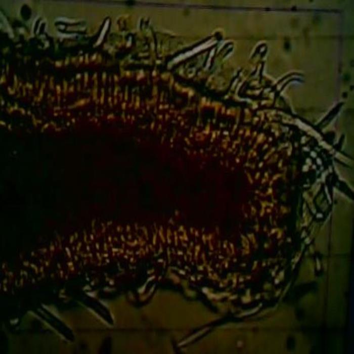
Microscopic characteristics
transverse section of the leaf exhibits its wing-like structure and it shows its isobilateral character where three layers of broad and short palisade cells occur beneath the upper epidermis and above the lower epidermis. Both epidermises bear conical, warty, and tapering covering trichomes that vary in length and thickness due to thick cell walls and narrow lumens. The epidermal cells are small in size and they are oval to round in shape, and they are covered with thick striated cuticle. The spongy mesophyll cells are oblong and somewhat distorted in shape. They are of variable sizes having thick walls, and most of them contain yellowish brown-coloured materials. Those that surround the vascular tissues are almost devoid of these coloured materials, and the majority of other spongy cells are compressed. The zone of palisade tissues above the lower epidermis is interrupted at the midrib zone and it is surrounded by distorted spongy cells with grey colour. The stem and branches are silvery-white with a pink tint. They bear stiff covering trichomes and leaves or leaves remaining are found at their numerous internodes. Falling leaves leave dark brown scars. The branches are brittle, and their outer layer separate on breaking. A surface view of the stem shows that the epidermis consists of colorless polygonal to almost rectangular cells that bear conical, tapering, and warty covering trichomes. A transverse section of the stem exhibits a circular outline. The epidermis consists of small cells, which are almost square, and it bears numerous trichomes, most of which are short with thick walls and narrow lumens. The epidermis is underlain by 5-6 layers of greyish, polygonal, or oblong cortical parenchyma cells, although the majority is distorted and compressed. These are underlain by a few layers of brown, polygonal cells having the characteristics of bark cells. These are followed by several layers of compressed cells that surround a large zone of heavily lignified vascular tissues. The vascular tissues surround oblong cells of the pith that have thick cell walls. Some of these cells contain brownish materials, but at the center of the pith, cells separate from each other, resulting in large hollow areas.
Powdered plant material
The material consists of the pounded aerial parts. It is a brownish, heterogeneous coarse powder with a violet tint, and it includes whitish longitudinal tissues. It has a pleasant straw-like odour with slight aroma but it is tasteless. Microscopically, the powder shows many free conical, warty, and tapering covering trichomes of various lengths and sizes; the relatively smaller ones belong to leaves while the significantly large ones are from stem and branches. The powder also shows light orange-brown fragments of the leaf with compact endings of palisade cells rounded in outlines; some of these fragments bear many covering trichomes. There are also many gray or grayish-brown fragments of fibro-vascular tissues of branches, some are thick and tightly packed, in addition to many fragments of light brown bark cells, polygonal or almost square in outlines.
Parts studied
leaf and stem
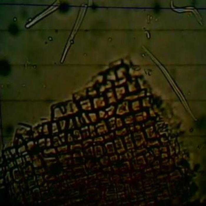
A) TS of leaf
B) Surface view of stem
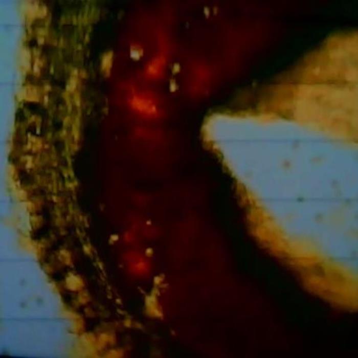
C) TS of the stem
- A. TS of a portion of the leaf near the margin of the lamina with the epidermises bearing conical warty trichomes. Three layers of the palisade tissues between the upper epidermis. At the center is the spongy mesophyll of which contain yellowish-brown materials.
- B. Surface view of a portion of the stem showing the almost rectangular, colorless epidermal cells with thick cell walls. The photo also shows some detached epidermal tapering covering trichomes.
- C. A transverse section of a representative portion of the stem showing different layers and zones: the small epidermal cells, layers of grayish cortical cells, layers of brownish polygonal cortical cells, layers of compressed cells surrounding a broad zone of heavily lignified vascular tissues then the pith cells part of which separate give a sizeable hollow area.
Chemical constituents
Beta acetoxy isovaleryl alkanin, beta dimethyl acryl shikonin (Salam, 1981). Benzoyl shikonin; beta-sitosterol, polyhydroxyterpine, kaempferol 4’-methyl ester-3-o-glucoside (Khafagy. 1981).
The following chemical studies have been carried out on the aerial part of the plant Maltkiopsis ciliata (Quality Control methods, 1998; Evans, 1996; ZCHRTM unpublished work):
Physicochemical constituents
Loss of weight in drying at 105°C : 11.40
Absolute alcohol solubility : 1.60
Water solubility : 12.00
Successive extractives (%)
Petroleum ether (60-80) °C : 1.00
Chloroform : 0.70
Absolute alcohol : 1.60
Ash values (%)
Total ash : 17.10
Water soluble ash : 5.10
Acid insoluble ash (10% HCl) : 1.60
pH values (aqueous solution)
pH of 1% solution : 8.362-8.396
pH of 10% solution : 7.960-7.970
Elemental analyses
Ash values (British Herbal Pharmacopeia)
Assay and identification of element (AOAC International)
|
Apparatus |
AA-6800 Shimadzu-Flame method |
||||
|
Element |
Std. conc. µg/ml(ppm) |
Sample conc. mg/ml |
Sample absorbance |
Actual conc.mg/ml |
Actual conc. (%) |
|
Cr |
1, 2, 4 |
10 |
0.0028 |
0.00456 |
0.000456 |
|
Zn |
0.25, 0.5, 1 |
10 |
0.0001 |
<0.0025 |
<0.0002 |
|
Cu |
1, 2, 4 |
10 |
0.0046 |
0.00554 |
0.000554 |
|
Fe |
1, 2, 4 |
0.909 |
0.0207 |
0.41976 |
0.041976 |
|
K |
1, 2, 4 |
0.909 |
0.2022 |
1.5423 |
0.15423 |
|
Pb |
1،2،4 |
10 |
0.0000 |
<0.001 |
<0.0001 |
|
Cd |
0.125, 0.25, 0.5 |
10 |
0.0000 |
<0.0001 |
<0.00001 |
|
Ca |
5, 10, 20 |
0.0826 |
0.1550 |
49.62819 |
4.96289 |
|
Mg |
0.25, 0.5, 1 |
0.0826 |
0.7977 |
8.21227 |
0.821227 |
|
Na |
1, 2, 4 |
0.909 |
0.1683 |
0.35002 |
0.035002 |
UV Spectral studies
|
Ultraviolet Spectrum (USP) |
||||
|
Apparatus |
Beckman DU 520 general purpose UV/VIS spectrophotometer. |
|||
|
Sample conc. (mg / ml) |
Solvent |
λ max (nm) |
λ min (nm) |
Abs.( λ max - λ min) |
|
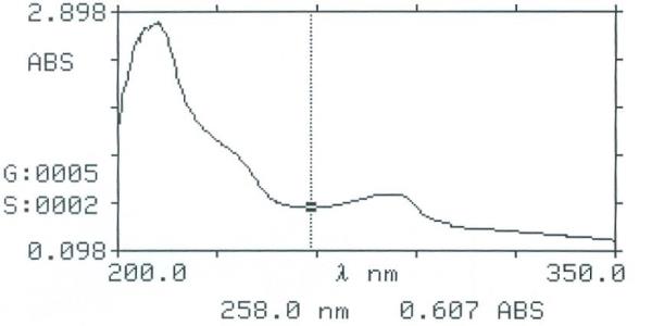
0.5481 |
Intestinal Fluid simulated without pancreatic pH=7.50.1 |
212.5 281 |
258 |
2.771 0.757- 0.607 |
|
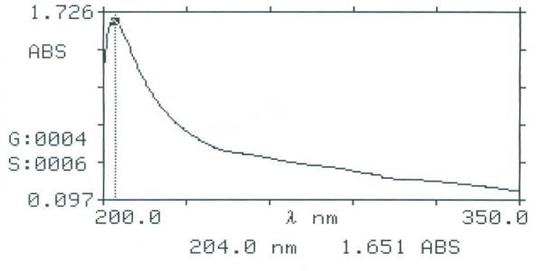
0.838 |
Gastric Fluid simulated without pepsin pH =1.20.1 |
204 |
|
1.651
|
Intestinal Fluid simulated without pancreatic
Gastric fluid simulated without pepsin
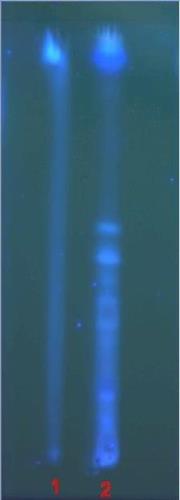
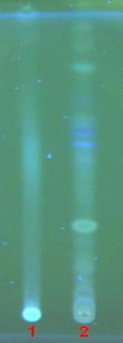
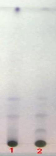
Chromatographical Studies
Thin layer chromatography (TLC): Wagner and Bladt, 1996

A
B
C
D
TLC fingerprint of Petroleum ether 60-80°C (track 1) and Methanol extract (track 2)
|
Mobile phase Fig. |
A&C |
: |
Toluene, ethyl formate, formic acid (5:4:1) |
|
|
B |
: |
Ethyl acetate, methanol, water (100:13.5:10) |
|
|
D |
: |
Toluene, ethyl acetate (93:7) |
|
Detection |
A |
: |
UV 254nm |
|
|
B&C |
: |
UV 366nm |
|
Derivatization |
D |
: |
Vanillin-Sulphuric acid-vis |
Toxicological and pharmacological Studies
Information contained in the literature about the plant:
Moltkiopsis sp. is a desert plant spreading from Morocco in North Africa to Saudi Arabia. It is palatable to camels. It is one the plants that was not studied for its pharmacological properties.
In highly polluted regions, ambient O3 pollution leads to a significant decrease in total sugars, pigments, and antioxidant enzymes activity in cultivated plant species. The results also showed that Bougainvillea spp. was more sensitive plant to O3 pollution compared to other cultivated plant species, while non-cultivated Moltkiopsis ciliata has more resistance than other plants. This investigation concluded that ozone pollution is responsible for the plant damage in industrial cities of KSA. (Akram, 2010)
The results on the pharmacological and Toxicological studies carried out at ZCHRTM labs. using the aqueous extract of Moltkiopsis ciliata (Derelanko 2002; Han, 2003) are shown in the following table:
|
ACTIVITY |
RESULTS |
|||
|
Strong |
Moderate |
Mild |
Negative |
|
|
Analgesic (Hot plate & Writhing) |
|
|
√ |
|
|
Anti-inflammatory (Paw edema) |
|
|
|
√ |
|
Effect on rabbit jejunum |
|
|
√ |
|
|
Effect on rat fundus strip |
|
|
√ |
|
|
Effect on detrusor muscle |
|
|
√ |
|
|
Effect on Guinea pig ileum |
|
|
|
√ |
|
Effect on right rat atria |
|
|
√ |
|
|
Anesthetized rat (BP & HR) |
|
|
√ |
|
|
Effect on thrombin time ↓ |
√ |
|
|
|
|
Biochemical studies |
|
|
|
√ |
|
Hematological studies |
|
|
|
√ |
|
Locomotor activity test ↓ |
|
|
√ |
|
|
Motor co-ordination (grip strength & motor activity) ↑ |
|
√ |
|
|
|
Rectal temperature |
|
|
|
√ |
|
Body weight |
|
|
|
√ |
|
Mortality |
|
|
|
√ |
Summary of the results
Both 70%alcohol and the water extracts showed significant decrease in prothrombin time. Spasmogenic activity of the plant indicates the purgative nature of the plant extract in relieving constipation and gastrointestinal spasms. The plant extract causes very mild relaxation indicating mild anti urolithic effect.
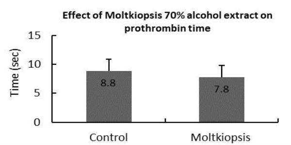
Effect on prothrombin time
Antimicrobial activity
The aqueous extract of the whole plant did not show antimicrobial activity against Mycobacterium smegmatis, C. tropicalis, different strains of Staphylococcus aureus (Including ATCC 257) as well as strains of Methicillin-Resistant Staphylococcus aureus, different strains of E. coli (Including ATCC UN 109), different strains of ESBL-producing K. pneumonia, Pseudomonas aeruginosa, and E. coli.
References
- Akram A. Ali and Mohammed N. El-Yemeni Atmospheric Air Pollution Effects on an Applied Sciences، 4(6): 1251-1263،2010.
- James P. Mandaville. Flora of Eastern Saudi Arabia. The Gresham Press، London. p.254، 1990.
- Jonbloed، M.V.، Feulner، G. R.، Boer، B. & Western، A. R. The comprehensive Guide to the Wild Flowers of the United Arab Emirates، Erwda، Abu Dhabi، U.A.E. (2003).
- Loutfy Boulos. Medicinal plants of North Africa، Reference Publications، USA، p.37، 1983.
- Mandaville J. P.، Flora of Eastern Saudi Arabia. Kegan Paul International، Riyadh، Saudi Arabia(1990).، Some Exhibited Plants at Aljubail Industrial City، KSA. Australian Journal of Basic
- Western، A. R. The Flora of United Arab Emirates. An introduction –Al Ain، (1986).
- Western، A. R. The Flora of United Arab Emirates. An introduction. Publications of the U.A.E. University (1989).